What Is a Brain AVM?

A brain arteriovenous malformation (AVM) is an abnormal tangle of blood vessels in which arteries connect directly to veins without the normal intervening capillary network. This creates a high-flow shunt that diverts blood away from normal brain tissue and places abnormal stress on the vessel walls, creating a risk of hemorrhage (bleeding), seizures, headaches, and progressive neurological deficits. Brain AVMs are believed to be congenital (present from birth) but may not produce symptoms until adolescence or adulthood. They are relatively rare, occurring in approximately 1 in 2,000 to 3,000 people, but they represent a significant cause of hemorrhagic stroke in young adults

Symptoms

  • Hemorrhage (brain bleeding) — the most common and most dangerous presentation, occurring in approximately 2-4% of AVM patients per year
  • Seizures — particularly common with superficial cortical AVMs
  • Headaches — which may be severe or atypical
  • Progressive neurological deficits — caused by “steal” of blood flow from surrounding normal brain
  • Pulsatile tinnitus — a rhythmic whooshing sound in the ear, when the AVM involves vessels near the skull base

Diagnosis & Grading

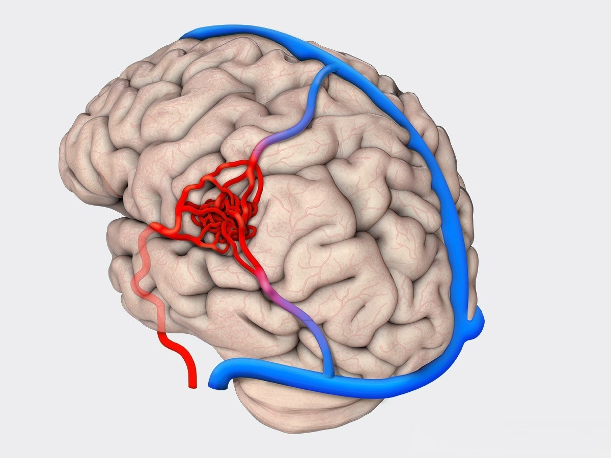
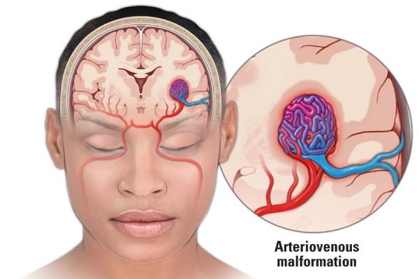
AVMs are evaluated with a combination of imaging studies:

  • MRI: Identifies the AVM location, size, and relationship to eloquent brain regions (areas controlling speech, movement, vision).
  • Catheter Angiography: The gold standard for characterizing AVM angioarchitecture—the detailed pattern of feeding arteries, nidus (the tangle itself), and draining veins. This information is essential for treatment planning.

AVMs are graded using the Spetzler-Martin scale (Grades I–V), which considers size, eloquence of adjacent brain, and pattern of venous drainage to estimate surgical risk. Dr. Choudhri is a contributor to the international MISTA Consortium, one of the world’s largest multicenter AVM research collaborations, with multiple publications analyzing treatment outcomes across all Spetzler-Martin grades.

Treatment Options

AVM treatment is often multimodal—meaning the best outcomes frequently come from combining two or more approaches. The treatment strategy is individualized based on the AVM’s grade, location, hemorrhage history, and patient factors.

Microsurgical Resection

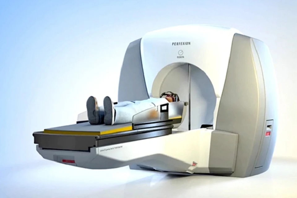
Complete surgical removal of the AVM nidus is the definitive cure and remains the gold standard for low-to-moderate grade AVMs (Spetzler-Martin I–III). When successful, microsurgery provides immediate elimination of the hemorrhage risk. Dr. Choudhri employs advanced techniques including intraoperative angiography (performed through transradial access), neuronavigation, neurophysiological monitoring, and fluorescence guidance to ensure complete resection while preserving normal brain function.

Endovascular Embolization

Catheter-based delivery of liquid embolic agents (such as Onyx) into the AVM feeding arteries to reduce blood flow through the lesion. Embolization is most commonly used as a preoperative adjunct to surgery—shrinking the AVM and reducing its blood flow to make surgical resection safer—or to target specific high-risk features such as intranidal aneurysms.

Dr. Choudhri’s MISTA Consortium research includes landmark studies on the safety and efficacy of preoperative embolization in AVMs, including analysis of outcomes in AVMs with perinidal aneurysms and single draining veins.

Stereotactic Radiosurgery (Gamma Knife / CyberKnife)

Focused radiation delivered precisely to the AVM nidus causes the abnormal vessels to gradually thicken and close over a period of 2–3 years. Radiosurgery is most effective for small AVMs (less than 3 cm) and is often used for deep-seated lesions in eloquent brain regions where surgery carries higher risk. Dr. Choudhri is certified in both Gamma Knife and CyberKnife radiosurgery.

Observation

Some AVMs—particularly those that are deep-seated, in critical brain regions, and have not hemorrhaged—may be most safely managed with careful observation and serial imaging. The decision not to treat is itself an active treatment decision that requires expert judgment.

Dr. Choudhri's AVM Expertise

Dr. Choudhri trained under Dr. Michael Lawton at UCSF, widely considered the leading AVM microsurgeon in the world. He has published extensively on AVM management as part of the international MISTA Consortium and has experience with the full spectrum of AVM grades and treatment modalities including hybrid procedures combining embolization and microsurgery in a single session.
×