What Is a Dural Arteriovenous Fistula?

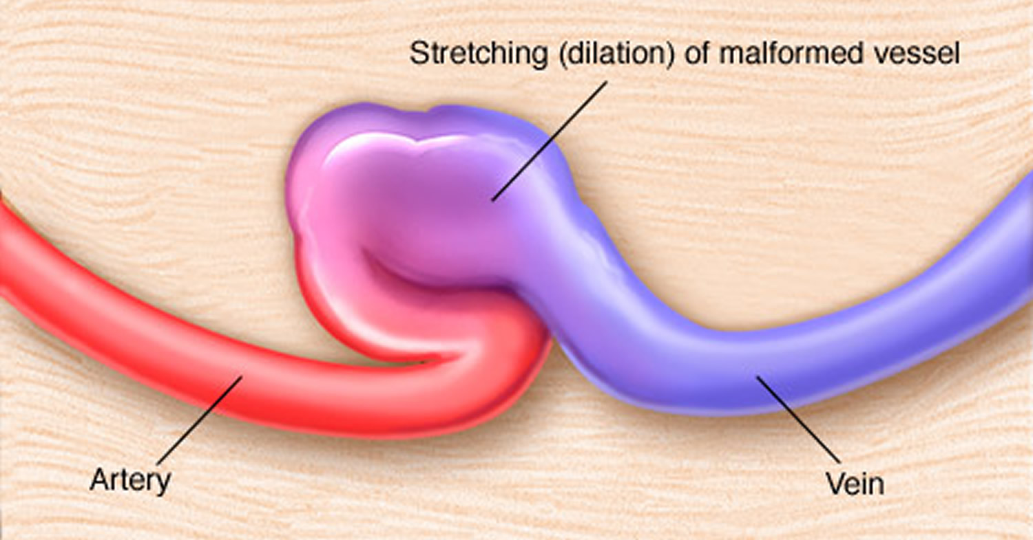
A dural arteriovenous fistula (dAVF) is an abnormal direct connection between arteries and veins within the dura mater—the tough membrane covering the brain and spinal cord. Unlike AVMs, which are congenital, dAVFs are typically acquired conditions that develop in response to venous sinus thrombosis, trauma, surgery, or infection.
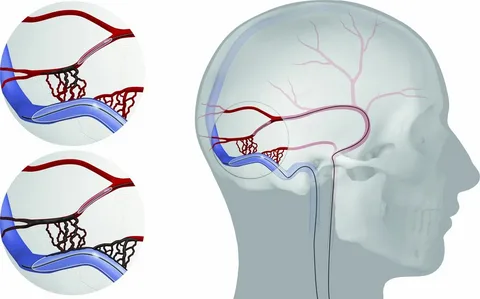
dAVFs are classified using the Cognard or Borden grading systems based on their venous drainage pattern, which determines their clinical risk. Fistulas draining into venous sinuses with normal flow direction (Cognard Type I) carry minimal risk, while those with cortical venous reflux (Cognard Types IIb–V) carry a significant risk of hemorrhage, venous infarction, and progressive neurological deterioration.

Symptoms

  • Pulsatile tinnitus: A rhythmic whooshing sound in the ear synchronized with the heartbeat—the most common presenting symptom of dAVFs near the transverse and sigmoid sinuses. Many patients see multiple doctors before the diagnosis is made.
  • Headaches
  • Visual disturbances or eye swelling (especially with cavernous sinus dAVFs)
  • Progressive cognitive decline or confusion (when cortical venous hypertension affects brain drainage)
  • Thalamic edema causing memory impairment or altered consciousness
  • Hemorrhage (brain bleeding)—the most feared complication of high-grade fistulas
  • Myelopathy (spinal cord dysfunction)—with spinal dAVFs
  •  

Diagnosis

  • MRI / MRA: Can show indirect signs of a dAVF including prominent vessels, edema, or abnormal flow voids. However, small fistulas can be missed.
  • CT Angiography: Useful for initial evaluation and identifying the fistula site.
  • Catheter Angiography: The definitive diagnostic test. Provides detailed characterization of feeding arteries, fistula point, and drainage pattern. Essential for treatment planning.
  • Balloon-Assisted Retrograde Cerebral Phlebography: An advanced diagnostic technique pioneered in Dr. Choudhri’s practice for superior visualization of cerebral venous anatomy, particularly in complex skull base venous pathology. This technique has been presented at SNIS and NASBS meetings.

Treatment Options

Endovascular Embolization

The first-line treatment for most dAVFs. A microcatheter is navigated through the arterial system (or in some cases the venous system) to the fistula point, and liquid embolic agents or coils are used to permanently close the abnormal connection. Cure rates for endovascular treatment of dAVFs are high—in many cases exceeding 80–90%. Dr. Choudhri’s approach emphasizes transradial (wrist) access when possible and utilizes the full range of embolic materials including Onyx, coils, and newer liquid embolics to achieve complete fistula obliteration.

Microsurgical Disconnection

When endovascular treatment is not feasible or does not achieve complete cure, open surgical disconnection of the draining vein(s) at the fistula point provides definitive treatment. This is particularly relevant for certain tentorial dAVFs and complex fistulas with multiple arterial feeders. Dr. Choudhri has published detailed case series on the microsurgical treatment of tentorial dural fistulas causing thalamic venous hypertension.

Combined Hybrid Approach

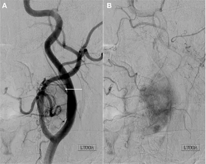
Some complex dAVFs benefit from a combined approach—embolization to reduce flow through the fistula followed by microsurgical disconnection—performed in a hybrid operating room that combines the capabilities of an angiography suite with a neurosurgical operating theater.

Many patients with pulsatile tinnitus caused by dAVFs have been told their symptoms are “just tinnitus” or have been given hearing aids without an adequate vascular workup. If you have pulsatile tinnitus that beats in time with your heartbeat, a thorough evaluation including advanced imaging can identify treatable causes. Dr. Choudhri established a dedicated multidisciplinary Pulsatile Tinnitus Program and has published diagnostic imaging algorithms for this condition.

×