Understanding Cerebral Venous Disorders

The venous system of the brain—the network of veins and sinuses that drains blood and cerebrospinal fluid from the brain back to the heart—has historically received far less attention than the arterial system. Yet disorders of cerebral venous drainage are increasingly recognized as important causes of debilitating symptoms including pulsatile tinnitus, headaches, vision loss, and elevated intracranial pressure. Dr. Choudhri’s clinical practice and research program place cerebral venous disease at the center of his expertise. He has pioneered advanced diagnostic techniques for venous imaging and has published some of the earliest outcome data for endovascular treatment of venous conditions.

Conditions Treated

Venous Sinus Stenosis

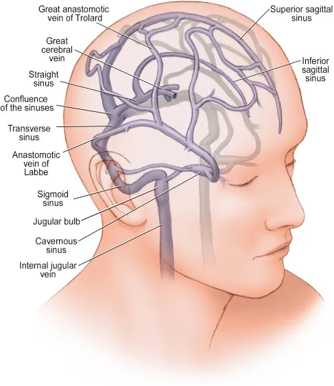
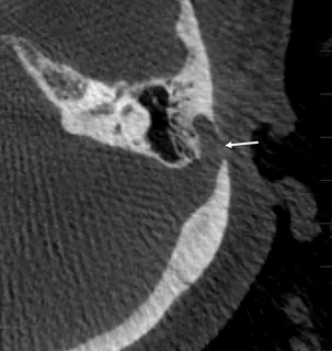
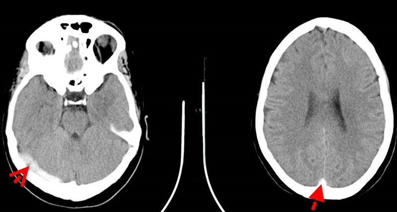
Narrowing of the major venous sinuses (most commonly the transverse and sigmoid sinuses) can impair drainage from the brain, leading to elevated intracranial pressure. Symptoms include headaches, pulsatile tinnitus, visual disturbances, and in severe cases, vision loss from papilledema. Venous sinus stenosis is frequently associated with idiopathic intracranial hypertension (IIH).

Idiopathic Intracranial Hypertension (IIH / Pseudotumor Cerebri)

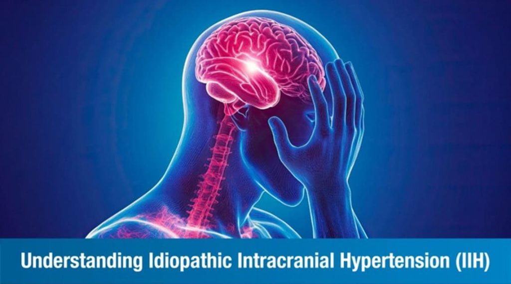
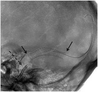
IIH is a condition of elevated pressure inside the skull without an identifiable structural cause. Patients experience severe headaches, pulsatile tinnitus, visual disturbances, and risk of permanent vision loss. IIH is increasingly understood to involve venous sinus stenosis as a contributing factor, and venous sinus stenting has emerged as an effective treatment option for selected patients.

Dr. Choudhri has published research on the role of stenting in venous sinus stenosis for IIH and is a contributor to the MPRESS trial, a PCORI-funded national study evaluating treatments for IIH. His team has also published on the emerging role of GLP-1 receptor agonists (such as semaglutide) in IIH management—an exciting new therapeutic avenue.

Venous Sinus Diverticula

A venous sinus diverticulum is a small outpouching of a venous sinus wall, frequently located near the ear at the junction of the transverse and sigmoid sinuses. These diverticula create turbulent blood flow that patients perceive as pulsatile tinnitus. While not dangerous, the symptoms can be severely debilitating and impact quality of life. Dr. Choudhri has published the clinical outcomes of endovascular treatment for venous sinus diverticula—one of the first outcome series in the literature for this condition—demonstrating high rates of symptom resolution.

Cerebral Venous aSinus Thrombosis (CVST)

Blood clot formation in the cerebral venous sinuses, which can cause headaches, seizures, stroke, and elevated intracranial pressure. Management involves anticoagulation and, in severe cases, endovascular thrombectomy or thrombolysis. Dr. Choudhri has published on both spontaneous venous thrombosis management and the rare complication of delayed venous thrombosis following fistula treatment.

Diagnostic Approach

  • MR Venography (MRV): Non-invasive evaluation of venous sinus anatomy and flow.
  • CT Venography (CTV): Detailed anatomical imaging of the venous sinuses and skull base.
  • Catheter Venography with Manometry: The gold standard for diagnosing venous stenosis. Pressure measurements are taken across the stenosis to confirm a significant pressure gradient, which guides treatment decisions.
  • Balloon-Assisted Retrograde Cerebral Phlebography: An advanced technique Dr. Choudhri pioneered for enhanced visualization of venous anatomy, particularly emissary veins and diverticular pathology. Presented at SNIS Cerebral Venous Summit.

Treatment Options

Venous Sinus Stenting

For patients with significant venous sinus stenosis and a documented pressure gradient, placement of a self-expanding stent within the stenotic sinus can restore normal venous drainage, reduce intracranial pressure, and resolve symptoms including pulsatile tinnitus and headache. Dr. Choudhri has published and presented on venous stenting outcomes and is a site principal investigator for the B-SILENT study evaluating the Sonorus Medical stent system

Endovascular Coiling of Venous Diverticula

Venous sinus diverticula causing pulsatile tinnitus can be treated by placing coils within the outpouching to eliminate the turbulent blood flow that produces the sound. This targeted endovascular approach can resolve the tinnitus while preserving normal sinus flow.

Medical Management

For IIH, initial management often includes weight loss counseling, acetazolamide (Diamox) to reduce CSF production, and emerging pharmacotherapy including GLP-1 receptor agonists. Surgical shunting (VP or LP shunt) may be considered for patients who fail medical therapy and are not candidates for stenting.

Upper Extremity Venous Access

Dr. Choudhri’s team has published the largest single-center experience using upper extremity venous access for neurointerventional procedures. This approach allows venous interventions—including sinus stenting and venous diagnostic procedures—to be performed through the arm rather than the groin, improving patient comfort and reducing access-site complications.

The Choudhri Lab’s research in cerebral venous disease represents one of the most active and productive programs in the country, with publications spanning diagnostic innovation, treatment outcomes, and novel therapeutic targets. Dr. Choudhri was invited faculty at the SNIS Cerebral Venous and CSF Disorders Summit (2024, 2025) and the New York Venous Symposium (2025).

×